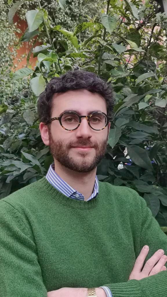
Alessandro Zampogna, Rome, Italy
Term of the Fellowship: 3.5. – 19.7.2021
Hosting department: Service de Neurologie, CHU Grenoble, Boulevard de la Chantourne, 38000 Grenoble, France
Supervisor: Prof. Elena Moro
My educational visit in the context of the EAN Clinical Fellowship Programme involved a period of 11 weeks at the Department of Neurology in the Centre Hospitalier Universitaire (CHU) of Grenoble, France, under the supervision of Prof. Elena Moro.
When applying to the EAN Clinical Fellowship Programme, I had no doubts that, based on my clinical and scientific interests in the field of movement disorders, Prof. Elena Moro and the CHU of Grenoble would have allowed a relevant educational period to me. Indeed, Prof. Moro is recognized as a leading expert in the field of movement disorders. Also, the CHU of Grenoble is the place where Deep Brain Stimulation (DBS) has been first developed and a renowned European centre for the management of movement disorders through advanced therapies.
Since the first day of my internship, I was introduced to all the members of Prof. Moro’s team and I was involved in the clinical activities at the CHU of Grenoble. More in detail, I followed the outpatient clinic implying medical consultations for patients suffering from several neurological conditions, including Parkinson’s disease, tremor, dystonia, Tourette syndrome, Huntington disease, and other rare movement disorders, such as paroxysmal dyskinesia and spinocerebellar ataxias. Attending the outpatient clinic allowed me to enhance my clinical skills in the diagnosis and therapeutic approach of patients affected by movement disorders. I have improved my knowledge in the management of patients with advanced Parkinson’s disease undergoing surgical or infusional therapies, including subcutaneous apomorphine and L-Dopa/Carbidopa intestinal gel infusions. I have been actively involved in all the procedures of DBS surgery, starting from the patient’s selection and education to the intraoperative and postoperative management. By attending all the procedures related to DBS surgery, I have appreciated the multidisciplinary and patient-centred approach of Prof. Moro’s team that in addition to the neurologists, involved a huge number of professional figures, including neurosurgeons, neuropsychologists, neurophysiologists, nurses and administrative staff.
Over the course of my educational period at the CHU of Grenoble, I also had the opportunity to train myself in botulin toxin injections, mainly for patients affected by dystonia. Prof. Moro contributed to enhancing my skills in injecting botulin toxin under both electromyography and ultrasound guidance. Furthermore, the possibility to join multidisciplinary consultations engaging geneticists and paediatricians helped me to better understand the diagnostic and therapeutic approaches of specific genetic disorders, such as myoclonus-dystonia, DYT1 generalised dystonia and GLUT-1 deficiency.
The wide spectrum of clinical activities that I have attended also included ward visits for patients admitted in the hospital, thus concerning the management of patients requiring more intensive medical care than those usually seen in the context of the outpatient clinic.
Despite the brief period of my internship at the CHU, Prof. Moro also gave me the opportunity to join the scientific activities of her research group by involving me in a new project regarding a large cohort of patients with Parkinson’s disease and DBS.
A final comment concerns the great cordiality and openness of the whole team headed by Prof. Moro, as well as the attractiveness offered by the city of Grenoble. During my educational period in Grenoble, there were several conviviality moments and sharing of extra-working experiences, including meals at the restaurant to taste some regional dishes. The international context of the CHU allowed me to meet colleagues from other European countries and enjoy together the magnificent landscapes of Grenoble. Grenoble is a quiet and functional city, ideal for those who love nature and mountain excursions, as well as cycling. Overall, at the end of my period at the CHU of Grenoble, I can certainly state that reality has matched my expectations. All the educational goals that I have set before my departure from Italy have been achieved completely. Accordingly, I would like to sincerely thank the European Academy of Neurology, Prof. Moro and her whole team (with a special mention for Dr Anna Castrioto, Dr Valerie Fraix, Dr Sara Meoni and Dr Sina Potel) for giving me this great opportunity that contributed to my professional and personal growth.
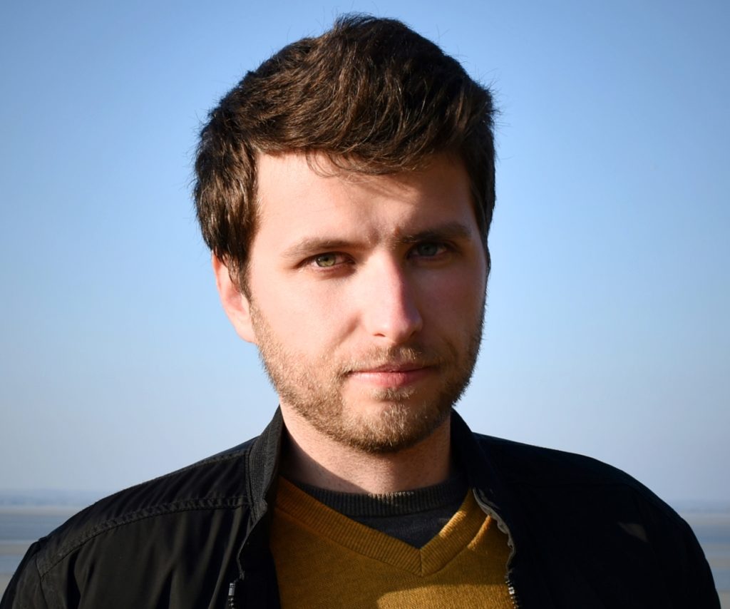
Dragos-Andrei Spinu, Iasi, Romania
Term of the Fellowship: 1.3.2021 – 30.4.2021
Hosting department: Van Gogh Epileptology Unit, Department of Neurology, Rennes University Hospital, France
Supervisor: Dr. Anca Nica
During my clinical fellowship, I attended the Van Gogh Epileptology Unit of the Rennes University Hospital in France, under the supervision of Dr. Anca Nica.
My primary objectives were to enrich my theoretical knowledge in the complex field of epilepsy and to get more more focused training on the electrophysiological aspects, both scalp and stereo-EEG. I also intended to gain more knowledge regarding the pharmacological, surgical or neuromodulatory treatment of different epilepsy types.
I had the opportunity to assist, work and get precious insights from Dr Anca Nica, Dr Mihai Maliia and Dr Arnaud Biraben during everyday clinical and teaching activities. They have a small but very cohesive Epilepsy Unit that also greatly benefits from astute and precise reports delivered by the unit’s neuropsychologist, Pascale Trebon and from the experience of three EEG technicians. Beside prolonged (1-3h) EEGs, they admit patients for long term (5days) scalp-EEG monitoring, as well as for intracranial stereo-EEG monitoring, in collaboration with the Neurosurgery department.
I was included in all the clinical activities and Dr Nica managed to arrange things so I could be exposed to all the aspects of epileptology. During the mornings, my activity consisted of seeing the patients admitted for the long term (5 days) EEG monitoring, watching and interpreting their video-EEG and discussing their case with the medical team. During the whole day I could also watch, interpret and discuss other EEGs with either Dr Maliia and Dr Nica. I increased my semiology knowledge, both clinical and electrical, through a series of video-EEG cases they have in their collection, some with puzzling semiology (especially the insular and cingular cases). I discovered quickly that even the apparently simpler cases or EEGs have in fact certain layers of complexity and various aspects to take into consideration. I could participate in their outpatient and day-hospital consultations, to see their approaches regarding pharmacological treatment regimens of focal or generalized epilepsies in various situations, as most patients had refractory epilepsy. I learnt the indications for a Vagus Nerve Stimulator and how to program the device’s settings. Beside studying the patients’ EEG, I appreciated the time spent to interpret and discuss many MRIs with either clearcut or just fine details of different structural abnormalities associated with epilepsy (mostly hippocampal sclerosis and different malformations of cortical development). Every day I was learning something new and I was motivated to read to further my understanding.
Every Friday morning there was a meeting with the epileptologists, pediatric neurologists, a neurologist specialized in genetics, and neurologists from other hospitals in Brittany where difficult clinical cases were discussed, mostly refractory epilepsy cases where either neuromodulatory or surgical treatment was to be proposed, or further exploration with intracranial electrodes. One of the most critical aspects of these meetings was the decision to implant depth electrodes for SEEG monitoring and choosing the locations for implantation, according to the electro-anatomo-clinical hypothesis.
The epileptologists’ collaboration with the Functional Neurosurgery department was excellent, no doubt one of the reasons for the patients’ good outcome. I was able to assist in the planning of implantation of SEEG electrodes, based on the electroclinical hypothesis and fine-tuned taking into consideration the MRI anatomy and vasculature. I have joined Dr Nica or Dr Maliia several times in the operating room, during the implantation of a new VNS electrode or battery replacement and I was taught how to program the device. I also had the chance to assist at a “classic” anterior temporal lobectomy for mesial temporal lobe epilepsy with hippocampal sclerosis and one frontal (SMA) cortectomy for frontal epilepsy due to focal cortical dysplasia, the latter allowing me to witness the use of electrocorticography to improve the limits of resection. On two occasions I could assist in the minimally invasive implantation of depth electrodes for the SEEG exploration, much aided by the neuronavigation system and the robotic stereotactic assistance. Needless to say, the OR is astounding.
Every Wednesday afternoon I participated in teaching courses or clinical presentations for the interns of the neurology department and there have also been 2 very valuable hands-on EEG courses organised by Dr Nica.
I was exposed for the first time to reading stereo-EEG, and it took me some time to get used to the type of signal, montage, normal appearing EEG patterns in different regions and what is defined as abnormal during seizures and in the interictal state. A two-months fellowship is by no means sufficient to dwell inside its complexity, but I had the opportunity to see and discuss SEEGs from patients implanted in various cerebral localisations. Another totally new aspect that I could witness was the electrical stimulation used in these patients for producing seizures in order to delineate the epileptogenic network, and for brain mapping of eloquent areas.
Another particular aspect was the logistical possibility to perform a SPECT study in the immediate aftermath of a seizure. The radionuclide (Tc99m) had to be procured every morning from the Nuclear Medicine Lab, so that in case of a witnessed focal seizure, it could be injected during the first seconds in order to study the ictal cerebral blood flow distribution, compared to a baseline SPECT performed on the day of admission. Thus, I also learnt about the use of SPECT and SISCOM as ancillary testing in focal epilepsy. Because of the Covid19 regulatory measures, it was difficult to profit of everything Rennes had to offer. Nevertheless, before the interdiction of traveling in the month of April, I managed to visit some fantastic places on the northern and southern coast of Brittany, which truly is a wonderful region of France. I would like to thank the EAN for making this fellowship possible and Dr Nica, Dr Maliia and Dr Biraben for sharing their knowledge and experience with me.
